What is Morton's Neuroma Surgery?
Morton’s Neuroma is one of the most common reasons for pain on the ball of the foot. A neuroma is a benign thickening or swelling of a normal nerve in the forefoot. Morton’s Neuroma is usually located in front of the third and fourth toes but can also be present between the second and third toes.
Morton's Neuroma Symptoms
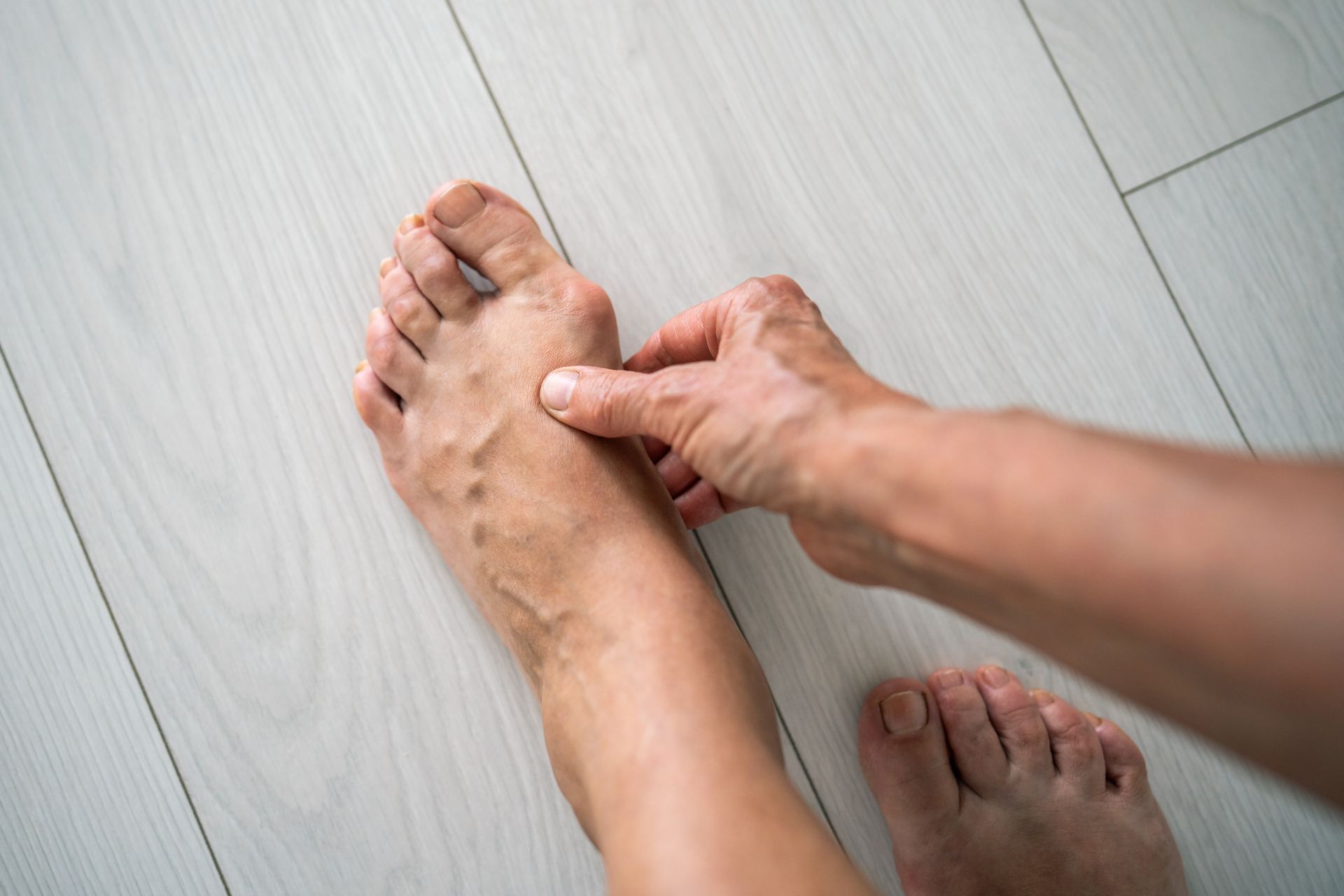
Symptoms of Morton’s Neuroma include pain, burning, tingling and numbness between the affected toes. Pain is usually caused by high-heeled, pointy-toed or narrow shoes and by activities that place added pressure on the ball of the foot.
Diagnosing Morton's Neuroma
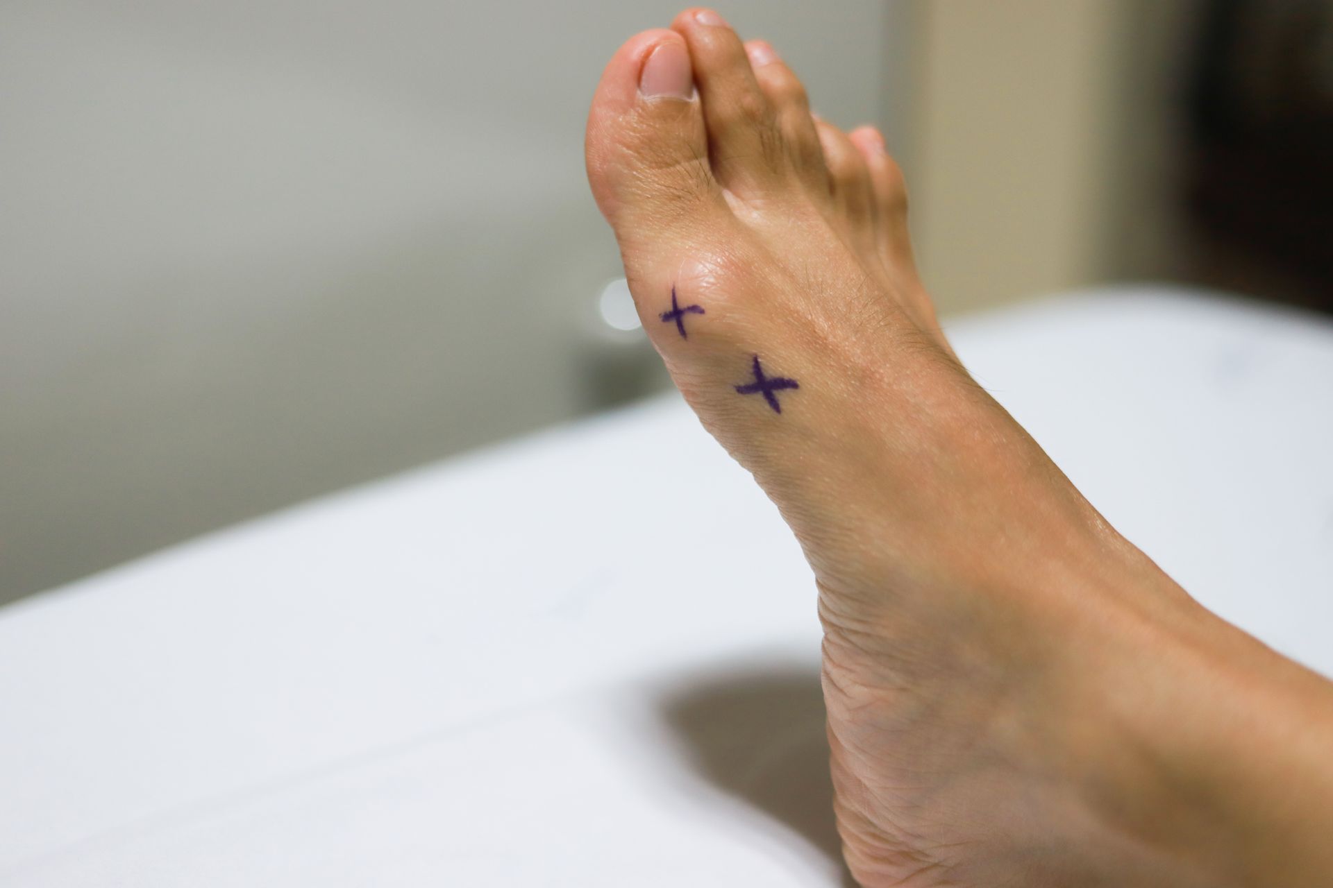
Diagnosing Morton’s Neuroma involves a detailed history and a physical examination. A test called Mulder sign helps diagnose neuroma. A foot specialist will push up on the symptomatic area with one thumb and squeeze the forefoot from side to side. A positive Mulder sign elicits a palpable click and reproduces the patient’s symptoms. X-rays do not show Morton’s Neuroma but can help rule out other diagnoses such as arthritis or stress fracture. In difficult to diagnose cases, an MRI can also be helpful.
Non-surgical Treatment of Morton's Neuroma
Most patients suffering from Morton’s Neuroma have good results with non-surgical treatments . These include injections, medications, pads, cushions, shoe inserts and, in some instances, physical therapy and walking boot immobilization. When non-surgical treatment fails, surgery to remove the Morton’s Neuroma yields good results.
Surgical Treatment for Morton's Neuroma
Surgical treatment for a painful Morton’s Neuroma involves a neurectomy or removal of the painful portion of nerve. A Morton’s Neuroma is one of the only nerves in the body that is routinely excised. There are two basic surgical approaches to removing a Morton’s Neuroma based on making an incision on the top or bottom of the foot.
Incision on the
Dorsum (top) of the Foot
This is certainly the most common
way to perform Morton’s Neuroma surgery. It is an outpatient surgery done under
IV sedation or twilight sleep. The surgeon typically injects a local numbing
medicine similar to Novocaine into the surgical site. This helps reduce the
amount of anesthesia medicine required and keeps patients comfortable through
the first night. The surgery takes less than an hour, and patients go home the
same day.
During the procedure, a 3 to 4 cm incision is made on top of the foot and directly over the neuroma. Meticulous dissection identifies the Morton’s Neuroma and all of its branches. The neuroma is carefully sectioned, removed from the foot and often sent to pathology to confirm diagnosis. The incision is closed with sutures.
Post-operative instructions will vary by physician. Generally, crutches are not needed for this procedure and patients do not need to be non-weightbearing. The first post-operative week is spent home, elevated and off the foot. Patients benefit from immobilization in a surgical shoe for 4 to 6 weeks and then transition to a regular shoe. Athletic activities can resume as early as 6 to 8 weeks post-op.
The advantages of this technique are the short recovery and good overall results. The primary disadvantage of this technique is the potential for recurrence, which is greater than when an incision is made on the bottom of the foot as described below.
Incision on the Bottom of the Foot (Plantar)
The basics of this approach are the same. It is an outpatient surgery, performed under IV sedation that takes less than an hour. Patients go home the same day and are given a local numbing medicine during the procedure to keep them comfortable the first night.
During this technique, a 4 to 5 cm incision is made on the bottom of the foot. Careful dissection enables the surgeon to excise a larger section of neuroma, reducing the rate of recurrence. In some instances, the end of the nerve is buried into muscle to reduce the rate of recurrence even more. The incision is closed with sutures.
The recovery from a plantar incision is different than a dorsal incision. This requires 2 to 4 weeks of non-weightbearing followed by 4 weeks in a walking boot or a surgical shoe. Painful scar is not a common complaint (less than 5%) but worries a lot of patients during pre-operative discussion. The lower recurrence rate combined with the more difficult recovery makes this technique useful for revision surgery, since recurrence with the first technique (top of the foot) is not common.